Gram-negative-staining Bacillaceae with thick cell wall and monoderm architecture uncover evolutionary diversity and challenge Gram-based classification
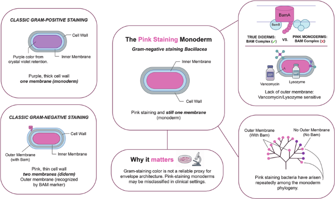
Gram staining has guided microbiology for over a century by coloring cells purple or pink, a read-out thought to distinguish monoderms (single membrane, thick peptidoglycan) from diderms (inner and outer membranes with thin wall). Here we show this rule fails repeatedly across Bacillaceae lineages historically deemed “Gram-positive”. By combining light and transmission-electron microscopy, antibiotic-sensitivity assays and comparative genomics across 57 strains, we identify “Gram-negative-staining monoderms” lacking outer membrane yet retaining thick peptidoglycan walls. These bacteria lack lipopolysaccharide- and β-barrel assembly-genes and remain highly susceptible to vancomycin and lysozyme (agents normally excluded by diderm envelopes), demonstrating functional monoderm status. Surprisingly, teichoic-acid biosynthetic pathways are patchily distributed and do not predict staining behavior. This discovery calls into question the textbook purple-or-pink dichotomy, decoupling stain color from membrane architecture. Clinically, misidentifying pink-staining Bacillaceae (including emerging pathogens such as Bacillus infantis) risks inappropriate therapy, whereas genome-guided diagnostics enable precise antibiotic stewardship. Genomic and microscopy evidence shows that the Bacillaceae, assumed to only stain Gram-positive, actually have some lineages staining Gram-negative, yet unexpectedly maintaining a thick wall and no outer membrane, challenging Gram staining assumptions.